Photodynamic Therapy
Photodynamic Therapy
Photodynamic therapy (PDT), sometimes called photochemotherapy, is a form of phototherapy using nontoxic light-sensitive compounds that are exposed selectively to light, whereupon they become toxic to targeted malignant and other diseased cells (phototoxicity). PDT has proven ability to kill microbial cells, including bacteria, fungi and viruses. PDT is popularly used in treating acne. It is used clinically to treat a wide range of medical conditions, including wet age-related macular degeneration and malignantcancers,[1] and is recognised as a treatment strategy which is both minimally invasive and minimally toxic.
Most modern PDT applications involve three key components:[1] a photosensitizer, a light source and tissue oxygen. The combination of these three components leads to the chemical destruction of any tissues which have both selectively taken up the photosensitizer and have been locally exposed to light. The wavelength of the light source needs to be appropriate for exciting the photosensitizer to produce reactive oxygen species. These reactive oxygen species generated through PDT are free radicals (Type I PDT) generated through electron abstraction or transfer from a substrate molecule and highly reactive state of oxygen known assinglet oxygen (Type II PDT). In understanding the mechanism of PDT it is important to distinguish it from other light-based and laser therapies such as laser wound healing and rejuvenation, or intense pulsed light hair removal, which do not require a photosensitizer.
Procedure
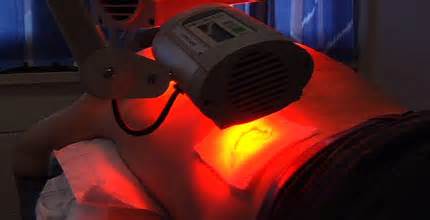
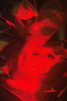
In order to achieve the selective destruction of the target area using PDT while leaving normal tissues untouched, either the photosensitizer can be applied locally to the target area, or photosensitive targets can be locally excited with light. For instance, in the treatment of skin conditions, including acne, psoriasis, and also skin cancers, the photosensitizer can be applied topically and locally excited by a light source. In the local treatment of internal tissues and cancers, after photosensitizers have been administered intravenously, light can be delivered to the target area using endoscopes and fiber optic catheters (see figure).
Photosensitizers can also target many viral and microbial species, including HIV and MRSA.[2] Using PDT, pathogens present in samples of blood and bone marrow can be decontaminated before the samples are used further for transfusions or transplants.[3] PDT can also eradicate a wide variety of pathogens of the skin and of the oral cavities. Given the seriousness that drug resistant pathogens have now become, there is increasing research into PDT as a new antimicrobial therapy.
Photosensitizers
In air and tissue, molecular oxygen occurs in a triplet state, whereas almost all other molecules are in a singlet state. Reactions between these are forbidden by quantum mechanics, thus oxygen is relatively non-reactive at physiological conditions. A photosensitizer is a chemical compound that can be promoted to an excited state upon absorption light and undergo intersystem crossing with oxygen to produce singlet oxygen. This species rapidly attacks any organic compounds it encounters, thus being highly cytotoxic. It is rapidly eliminated: in cells, the average lifetime is 3 µs.[5]
A wide array of photosensitizers for PDT exist. They can be divided into porphyrins, chlorophylls and dyes.[6] Some examples includeaminolevulinic acid (ALA), Silicon Phthalocyanine Pc 4, m-tetrahydroxyphenylchlorin (mTHPC), and mono-L-aspartyl chlorin e6 (NPe6).
Several photosensitizers are commercially available for clinical use, such as Allumera, Photofrin, Visudyne, Levulan, Foscan, Metvix,Hexvix, Cysview, and Laserphyrin, with others in development, e.g. Antrin, Photochlor, Photosens, Photrex, Lumacan, Cevira, Visonac,BF-200 ALA. Amphinex. Also Azadipyrromethenes.
Although these photosensitizers can be used for wildly different treatments, they all aim to achieve certain characteristics:
- High absorption at long wavelengths
- Tissue is much more transparent at longer wavelengths (~700–850 nm). Absorbing at longer wavelengths would allow the light to penetrate deeper, and allow the treatment of larger tumors.
- High singlet oxygen quantum yield
- Low photobleaching to prevent degradation of the photosensitizer[why?]
- Natural fluorescence
- Many optical dosimetry techniques, such as fluorescence spectroscopy, depend on the drug being naturally fluorescent
- Many optical dosimetry techniques, such as fluorescence spectroscopy, depend on the drug being naturally fluorescent
- High chemical stability
- Low dark toxicity
- The photosensitizer should not be harmful to the target tissue until the treatment beam is applied.
- Preferential uptake in target tissue
The major difference between different types of photosensitizers is in the parts of the cell that they target. Unlike in radiation therapy, where damage is done by targeting cell DNA, most photosensitizers target other cell structures. For example, mTHPC has been shown to localize in the nuclear envelope and do its damage there. In contrast, ALA has been found to localize in the mitochondria and Methylene Blue in the lysosomes.
To allow treatment of deeper tumours some researchers are using internal chemiluminescence to activate the photosensitiser.
PUVA therapy is using psoralen as photosensitiser and UVA ultraviolet as light source, but this form of therapy is usually classified as a separate form of therapy from photodynamic therapy.
Targeted PDT[edit]
Some photosensitisers naturally accumulate in the endothelial cells of vascular tissue allowing ‘vascular targeted’ PDT, but there is also research to target the photosensitiser to the tumour (usually by linking it to antibodies or antibody fragments). It is currently only in pre-clinical studies.[17][18]
Applications[edit]
Compared to normal tissues, most types of cancers are especially active in both the uptake and accumulation of photosensitizers agents, which makes cancers especially vulnerable to PDT.[19] Since photosensitizers can also have a high affinity for vascular endothelial cells.[20]
Usage in acne[edit]
PDT is currently in clinical trials to be used as a treatment for severe acne. Initial results have shown for it to be effective as a treatment only for severe acne,[21] though some question whether it is better than existing acne treatments. The treatment causes severe redness and moderate to severe pain and burning sensation. (see also: Levulan) A phase II trial, while it showed improvement occurred, failed to show improved response compared to the blue/violet light alone.
History
While the applicability and potential of PDT has been known for over a hundred years,[23] the development of modern PDT has been a gradual one, involving scientific progress in the fields of photobiology and cancer biology, as well as the development of modern photonic devices, such as lasers and LEDs.[24] It was John Toth as product manager for Cooper Medical Devices Corp/Cooper Lasersonics with early clinical argon dye lasers ca 1981 who acknowledged the “photodynamic chemical effect” of the therapy and wrote the first “white paper” branding the therapy as “Photodynamic Therapy” (PDT) to support efforts in setting up 10 clinical sites in Japan where the term “radiation” had negative connotations. PDT received even greater interest as result of Thomas Dougherty helping expand clinical trials and forming the International Photodynamic Association, in 1986.
In the 1990s the team of Polish professor Aleksander Sieroń developed the process and devices used today for PDT treatment, while improving the photosensitizer compound at the university of Bytom in Poland. This Polish team is considered at the cutting edge of research in this field, with many published articles and ongoing clinical trials.
PDT in ancient medicine
The earliest recorded treatments that exploited a photosensitizer and a light source, in this case sunlight, for medical effect can be found in ancient Egyptian and Indian sources. Annals over 3000 years old report the use of topically applied vegetable and plant substances to produce photoreactions in skin and cause a repigmentation of depigimented skin lesions, as seen with vitiligo and leukoderma.
The photosensitizing agents used in these ancient therapies have been characterised with modern science as belonging to the psoralen family of chemicals. Psoralens are still in use today in PDT regimes to treat a variety of skin conditions, including vitiligo, psoriasis, neurodermatitis, eczema, cutaneous T-cell lymphoma and lichen ruber planus.
20th-century development of PDT
The first detailed scientific evidence that agents, photosensitive synthetic dyes, in combination with a light source and oxygen could have potential therapeutic effect was made at the turn of the 20th century in the laboratory of von Tappeiner in Munich, Germany. Historically this was a time when Germany was leading the world in the industrial synthesis of dyes.
While studying the effects of acridine on paramecia cultures, Oscar Raab, a student of von Tappeiner observed a toxic effect. Fortuitously Raab also observed that light was dependent for the killing of paramecia cultures to take place.[26] Subsequent work in the laboratory of von Tappeiner showed that oxygen was essential for the ‘photodynamic action’ – a term coined by von Tappeiner.[27]
With the discovery of photodynamic effects, von Tappeiner and colleagues went on to perform the first PDT trial in patients with skin carcinoma using the photosensitizer, eosin, Out of 6 patients with a facial basal cell carcinoma, treated with a 1% eosin solution and a long-term exposure either to sunlight or to arc-lamp light, 4 patients showed total tumour resolution and a relapse-free period of 12 months.
It was only much later, when Thomas Dougherty and co-workers[29] at Roswell Park Cancer Institute, Buffalo NY, clinically tested PDT again. In 1978, they published striking results in which they treated 113 cutaneous or subcutaneous malignant tumors and observed a total or partial resolution of 111 tumors.[30]
The active photosensitizer used in the clinical PDT trial by Dougherty was an agent called Haematoporphyrin Derivative (HpD), which was first characterised in 1960 by Lipson.[31]In his research, Lipson wanted to find a diagnostic agent suitable for the detection of tumours in patients. With the discovery of HpD, Lipson went onto pioneer the use of endoscopes and HpD fluorescence to detect tumours.[32]
As its name suggests, HpD is a porphyrin species derived from haematoporphyrin, Porphyrins have long been considered as suitable agents for tumour photodiagnosis and tumour PDT because cancerous cells exhibit a significantly greater uptake and affinity for porphyrins compared to normal quiescent tissues. This important observation, which underlies the success of PDT to treat cancers, had been established by a number of scientific researchers prior to the discoveries made by Lipson. In 1924, Policard first revealed the diagnostic capabilities of hematoporphyrin fluorescence when he observed that ultraviolet radiation excited red fluorescence in the sarcomas of laboratory rats.[33] Policard hypothesized at the time that the fluorescence was associated with endogenous hematoporphyrin accumulation. In 1948, Figge with co-workers[34] showed on laboratory animals that porphyrins exhibit a preferential affinity to rapidly dividing cells, including malignant, embryonic, and regenerative cells, and because of this, they proposed that porphyrins should be used in the treatment of cancer. Subsequently many scientific authors have repeated the observation that cancerous cells naturally accumulate porphyrins and have characterised a number of mechanisms to explain it.
HpD, under the pharmaceutical name Photofrin, was the first PDT agent approved for clinical use in 1993 to treat a form of bladder cancer in Canada. Over the next decade, both PDT and the use of HpD received wider international attention and grew in their clinical use, and lead to the first PDT treatments to receive U.S. Food and Drug Administrationapproval. Additional organic dyes applicable to laser PDT are listed by Goldman.
Modern development of PDT in Russia
Of all the nations beginning to use PDT in the late 20th century, the Russians were the quickest to advance its use clinically and to make many developments. One early Russian development was a new photosensitizer called Photogem which, like HpD, was derived from haematoporphyrin in 1990 by Professor Andrey F. Mironov and coworkers in Moscow. Photogem was approved by the Ministry of Health of Russia and tested clinically from February 1992 to 1996. A pronounced therapeutic effect was observed in 91 percent of the 1500 patients that underwent PDT using Photogem, with 62 percent having a total tumor resolution. Of the remaining patients, a further 29 percent had a partial tumor resolution, where the tumour at least halved in size. In those patients that had been diagnosed early, 92 percent of the patients showed complete resolution of the tumour.
Around this time, Russian scientists also collaborated with NASA medical scientists who were looking at the use of LEDs as more suitable light sources, compared to lasers, for PDT applications.
Modern development of PDT in Asia
PDT has also seen considerably development in Asia. Since 1990, the Chinese have been developing specialist clinical expertise with PDT using their own domestically produced photosensitizers, derived from Haematoporphyrin, and light sources.[38] PDT in China is especially notable for the technical skill of specialists in effecting resolution of difficult to reach tumours .